Gallbladder Adenomyomatosis in Pediatrics: A Case Report
DOI:
https://doi.org/10.22516/25007440.1018Keywords:
Gallbladder, Cholecystitis, Pancreatitis, Laparoscopy, PediatricsAbstract
Objective: Gallbladder adenomyomatosis (ADM) is a rare disease in pediatrics, characterized by epithelial proliferation and muscle hypertrophy, associated with the formation of fistulous tracts, classically called Rokitansky-Aschoff sinuses. It is an anatomical and clinical entity that is difficult to diagnose. Ultrasound is the primary diagnostic tool, but ADM is confirmed by its characteristic histological findings.
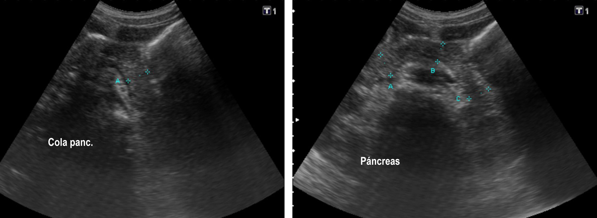
Clinical case: A 15-year-old adolescent with a previous diagnosis of congenital scoliosis and hemivertebra at T11-12 with secondary spinal cord compression and butterfly vertebrae at T5-L3 was admitted for surgical management of this entity. In the immediate postoperative period, band-like abdominal pain and elevated pancreatic enzymes were present, considering the initial diagnosis of acute pancreatitis. Imaging studies revealed a thickened gallbladder with increased size and findings compatible with gallbladder ADM. The patient showed improvement in symptoms after undergoing laparoscopic cholecystectomy. The diagnosis of gallbladder ADM was later confirmed by histology.
Conclusion: Gallbladder ADM is extremely rare in children; little is known about its pathogenesis and pathology. It is diagnosed mainly by ultrasound, which identifies hypertrophy of the muscular layer and the formation of fistulous tracts, known as Rokitansky-Aschoff sinuses.
Downloads
References
Burgos AM, Csendes A, Villanueva M, Cárdenas G, Narbona S, Caballero M, et al. Hallazgos clínicos e histopatológicos en pacientes con adenomiomatosis vesicular. Rev Cir. 2016;68(5) 363-367. https://doi.org/10.1016/j.rchic.2016.06.010
Zarate YA, Bosanko KA, Jarasvaraparn C, Vengoechea J, McDonough EM. Description of the first case of adenomyomatosis of the gallbladder in an infant. Case Rep Pediatr. 2014;2014:248369. https://doi.org/10.1155/2014/248369
Pasierbek M, Korlacki W, Grabowski A. Adenomiomatosis vesicular en un adolescente, una afección muy rara. Arch Argent Pediatr. 2020;118(1):43-7. https://doi.org/10.5546/aap.2020.e43
Pérez-Alonso AJ, Argote-Camacho ÁX, Rubio-López J, Olmo-Rivas D, Petrone P. Adenomiomatosis de la vía biliar, incidencia de 10 años y revisión de la bibliografía actual. Rev Colomb Cir. 2015;30(2) 112-118. https://doi.org/10.30944/20117582.319
Bonatti M, Vezzali N, Lombardo F, Ferro F, Zamboni G, Tauber M, et al. Gallbladder adenomyomatosis: imaging findings, tricks and pitfalls. Insights Imaging. 2017;8(2):243-253. https://doi.org/10.1007/s13244-017-0544-7
Parolini F, Indolfi G, Magne MG, Salemme M, Cheli M, Boroni G, et al. Adenomyomatosis of the gallbladder in childhood: A systematic review of the literature and an additional case report. World J Clin Pediatr. 2016; 5(2) 223-7. https://doi.org/10.5409/wjcp.v5.i2.223
Akçam M, Buyukyavuz I, Çiriş M, Eriş N. Adenomyomatosis of the gallbladder resembling honeycomb in a child. Eur J Pediatr. 2008;167(9):1079-1081. https://doi.org/10.1007/s00431-007-0623-8
Lam CM, Yuen AW, Wai AC, Leung RM, Lee AY, Ng KK, et al. Gallbladder cancer presenting with acute cholecystitis: a population-based study. Surg Endosc. 2005;19(5):697-701. https://doi.org/10.1007/s00464-004-9116-2
Sermon A, Himpens J, Leman G. Symptomatic adenomyomatosis of the gallbladder--report of a case. Acta Chir Belg. 2003;103(2):225-9. https://doi.org/10.1080/00015458.2003.11679412
Haradome H, Ichikawa T, Sou H, Yoshikawa T, Nakamura A, Araki T, et al. The pearl necklace sign: an imaging sign of adenomyomatosis of the gallbladder at MR cholangiopancreatography. Radiology. 2003;227(1):80-8. https://doi.org/10.1148/radiol.2271011378
Menocal N, Garrote A, García DA, Santos J. Vesícula biliar multiseptada: Una anomalía congénita infrecuente. Revista Chilena de Radiología. 2011;17(4):176-178. https://doi.org/10.4067/S0717-93082011000400006
Yu MH, Lee JY, Yoon JH, Baek JH, Han JK, Choi BI. Color Doppler twinkling artifacts from gallbladder adenomyomatosis with 1.8 MHz and 4.0 MHz color Doppler frequencies. Ultrasound Med Biol. 2012;38(7):1188-94. https://doi.org/10.1016/j.ultrasmedbio.2012.03.010
Cariati A, Cetta F. Rokitansky-Aschoff sinuses of the gallbladder are associated with black pigment gallstone formation: a scanning electron microscopy study. Ultrastruct Pathol. 2003;27(4):265-70. https://doi.org/10.1080/01913120309913
Downloads
Published
How to Cite
Issue
Section
License
Copyright (c) 2024 Revista colombiana de Gastroenterología

This work is licensed under a Creative Commons Attribution-NonCommercial-NoDerivatives 4.0 International License.
Aquellos autores/as que tengan publicaciones con esta revista, aceptan los términos siguientes:
Los autores/as ceden sus derechos de autor y garantizarán a la revista el derecho de primera publicación de su obra, el cuál estará simultáneamente sujeto a la Licencia de reconocimiento de Creative Commons que permite a terceros compartir la obra siempre que se indique su autor y su primera publicación en esta revista.
Los contenidos están protegidos bajo una licencia de Creative Commons Reconocimiento-NoComercial-SinObraDerivada 4.0 Internacional.

| Article metrics | |
|---|---|
| Abstract views | |
| Galley vies | |
| PDF Views | |
| HTML views | |
| Other views | |