Proximal esophageal stricture due to lymphocytic esophagitis: case report
DOI:
https://doi.org/10.22516/25007440.1521Keywords:
Esophagitis, dysphagia, esophageal strictureAbstract
Introduction: Lymphocytic esophagitis (LE) is an uncommon cause of dysphagia, characterized by peripapillary infiltration of intraepithelial lymphocytes. Its phenotypic presentation as a severe proximal esophageal stricture is exceptional.
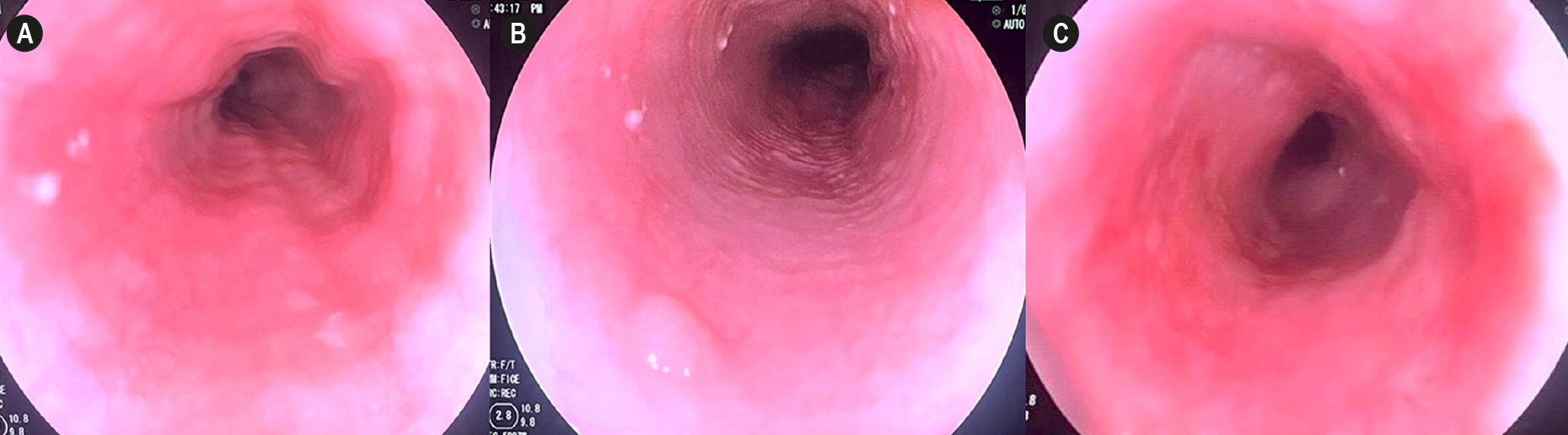
Case report: A 79-year-old man presented with progressive dysphagia over 11 months and a 9 kg weight loss. Endoscopy revealed a pinpoint cervical stricture. After an initial unsuccessful session, a second session of progressive pneumatic dilation successfully traversed the lesion, revealing "trachealized" mucosa with exudates. Histopathological examination of the biopsies showed basal hyperplasia and an infiltrate of 22 to 30 lymphocytes per high-power field in the peripapillary epithelium, without eosinophils, confirming LE. Treatment with omeprazole 20mg twice daily for 6 months achieved clinical and endoscopic resolution, without the need for repeat dilation.
Discussion: LE requires strict histopathological differentiation from eosinophilic esophagitis. In the absence of clinical guidelines, a stepwise empirical approach remains the current standard of care.
Conclusion: LE should be considered in the differential diagnosis of proximal esophageal stricture in older adults. Combined therapy with progressive pneumatic dilation and proton pump inhibitor has proven to be a safe and effective medium-term strategy.
Downloads
References
Rubio C, Sjodahl K, Lagergren J. Lymphocytic esophagitis: a histologic subset of chronic esophagitis. Am J Clin Pathol. 2006;125(3):432-37. https://doi.org/10.1309/7LA8LGY08UEM3H26
Rouphael C, Gordon IO, Thota PN. Lymphocytic esophagitis: still an enigma a decade later. World J Gastroenterol. 2017;23(6):949-56. https://doi.org/10.3748/wjg.v23.i6.949.
Visaggi P, Savarino E, Del Corso G, Herlihy N, Ghisa M, Dunn JM, et al. Clinical characteristics, endoscopic findings, and treatment outcomes in lymphocytic esophagitis compared with eosinophilic esophagitis. Am J Gastroenterol. 2025;120(2):469-72. https://doi.org/10.14309/ajg.0000000000003046
Haque S, Genta RM. Lymphocytic oesophagitis: clinicopathological aspects of an emerging condition. Gut. 2012;61(8):1108-14. https://doi.org/10.1136/gutjnl-2011-301014
Cohen S, Saxena A, Waljee AK, Piraka C, Purdy J, Appelman H, et al. Lymphocytic esophagitis: a diagnosis of increasing frequency. J Clin Gastroenterol. 2012;46(10):828-32. https://doi.org/10.1097/MCG.0b013e3182500de8
Zaver HB, Ghoz H, Malviya BJ, Brahmbhatt B, Palmer WC, Lacy BE, et al. Lymphocytic esophagitis: assessing risk factors and clinical outcomes. Dig Dis Sci. 2021;66(11):3976-84. https://doi.org/10.1007/s10620-020-06706-4
Hussein M, Mitchison M, Sweis R. Lymphocytic oesophagitis: diagnosis and management. Clin Med (Lond). 2023;23(6):540-44. https://doi.org/10.7861/clinmed.2023-0440
Reddy CA, McGowan E, Yadlapati R, Peterson K. AGA clinical practice update on esophageal dysfunction due to disordered immunity and infection: expert review. Clin Gastroenterol Hepatol. 2024;22(12):2378-87. https://doi.org/10.1016/j.cgh.2024.08.027
Habbal M, Scaffidi MA, Rumman A, Khan R, Ramaj M, Al-Mazroui A, et al. Clinical, endoscopic, and histologic characteristics of lymphocytic esophagitis: a systematic review. Esophagus. 2019;16(2):123-32. https://doi.org/10.1007/s10388-018-0649-1
Dakkak M, Bennett JR. A new dysphagia score with objective validation. J Clin Gastroenterol. 1992;14(2):99-100. https://doi.org/10.1097/00004836-199203000-00004
Greuter T, Straumann A, Fernandez-Marrero Y, Germic N, Hosseini A, Yousefi S, et al. Characterization of eosinophilic esophagitis variants by clinical, histological, and molecular analyses: a cross-sectional multi-center study. Allergy. 2022;77(8):2520-33. https://doi.org/10.1111/all.15233
Hendy PJ, Wong DS, Florin TH. Spontaneous oesophageal perforation: an unreported complication of lymphocytic oesophagitis. Gut. 2013;62(11):1668-69. https://doi.org/10.1136/gutjnl-2013-305455
Nguyen B, Cheng CF, Jowhari F. A systematic review of therapeutic options for lymphocytic esophagitis. Dis Esophagus. 2025;38(6):doaf112. https://doi.org/10.1093/dote/doaf112
Downloads
Published
How to Cite
Issue
Section
License
Copyright (c) 2026 Revista colombiana de Gastroenterología

This work is licensed under a Creative Commons Attribution-NonCommercial-NoDerivatives 4.0 International License.
Aquellos autores/as que tengan publicaciones con esta revista, aceptan los términos siguientes:
Los autores/as ceden sus derechos de autor y garantizarán a la revista el derecho de primera publicación de su obra, el cuál estará simultáneamente sujeto a la Licencia de reconocimiento de Creative Commons que permite a terceros compartir la obra siempre que se indique su autor y su primera publicación en esta revista.
Los contenidos están protegidos bajo una licencia de Creative Commons Reconocimiento-NoComercial-SinObraDerivada 4.0 Internacional.


| Article metrics | |
|---|---|
| Abstract views | 2 |
| Galley views | 0 |
| PDF Views | 0 |
| HTML views | 0 |
| Other views | 0 |