Endoscopic ultrasound-guided radiofrequency ablation in a pancreatic neuroendocrine tumor: expanding access to minimally invasive therapies
DOI:
https://doi.org/10.22516/25007440.1494Keywords:
Endoscopic ultrasonography, neuroendocrine tumor, radiofrequency ablationAbstract
Introduction: Pancreatic neuroendocrine tumors (pNETs) are uncommon neoplasms that originate in pancreatic islet cells and account for 1%-2% of all pancreatic tumors.
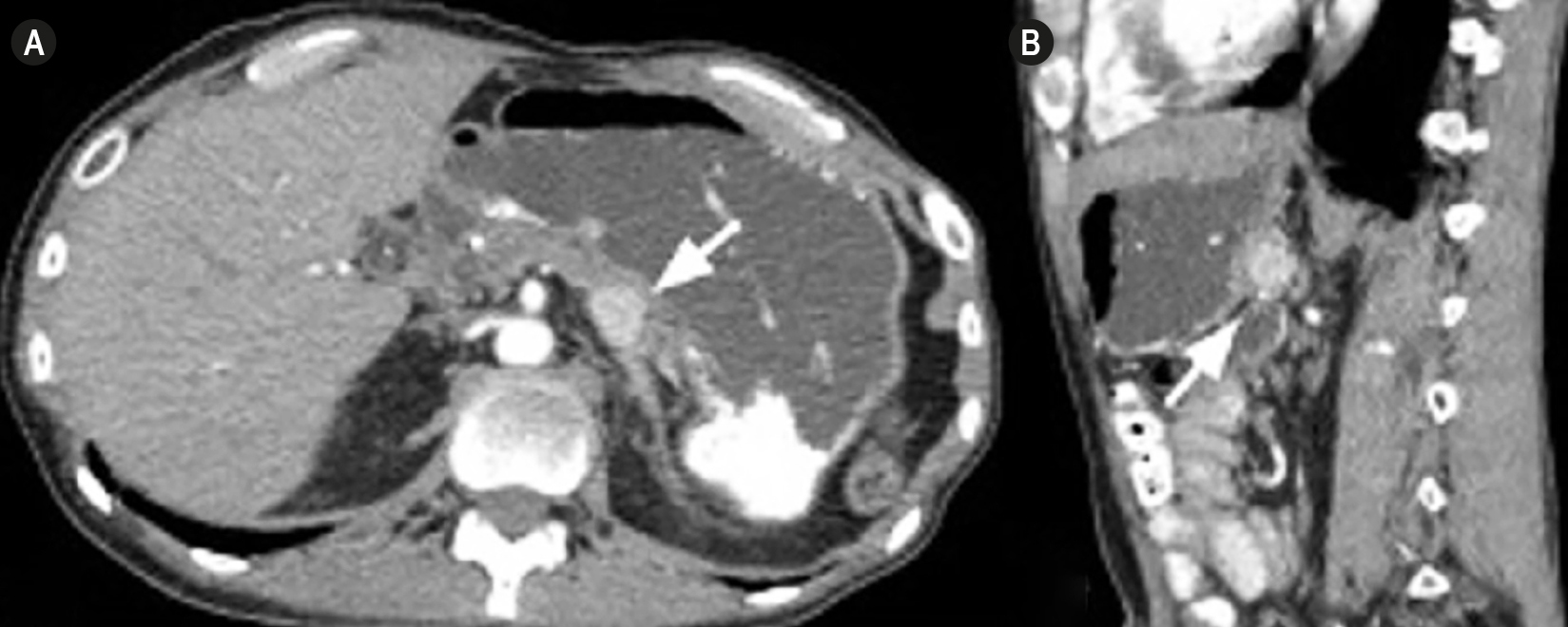
Case presentation: We present the case of a 47-year-old man with multiple pathological conditions and previous abdominal surgical interventions who sought medical care because of persistent elevation of liver enzymes. Magnetic resonance cholangiography showed an 18 mm solid lesion in the pancreatic body. Endoscopic ultrasonography (EUS) identified a well-defined, hypoechoic, hypervascular nodule. Fine-needle biopsy (FNB) confirmed a well-differentiated grade 1 pNET (Ki-67 1%). PET-CT with 68Ga-DOTA showed focal uptake consistent with a pNET. After a multidisciplinary discussion, the team chose EUS-guided radiofrequency ablation (EUS-RFA) because of the high surgical risk associated with the patient’s comorbidities and previous abdominal surgeries. Under general anesthesia, the team applied 30 W in continuous mode through a 19 G radiofrequency needle until achieving complete ablation.
Results: No immediate complications occurred. Computed tomography one week later confirmed complete tumor ablation without residual enhancement.
Conclusions: This case is consistent with international reports showing high efficacy and a low complication rate with EUS-RFA. However, the technique is not free of risks (pancreatitis, bleeding, perforation), which were not observed in this patient. As one of the first experiences in Latin America and, to the best of current knowledge, in Colombia, this case highlights that EUS-RFA is a promising minimally invasive alternative for selected pNETs. Regional cohorts are required to characterize risks and population-specific features, and comparative evaluations against active surveillance and surgery are justified.
Downloads
References
Ohmoto A, Rokutan H, Yachida S. Pancreatic neuroendocrine neoplasms: Basic biology, current treatment strategies and prospects for the future. Int J Mol Sci 2017;18(1):143. https://doi.org/10.3390/ijms18010143
Kos-Kudła B, Castaño JP, Denecke T, Grande E, Kjaer A, Koumarianou A, et al. European Neuroendocrine Tumour Society (ENETS) 2023 guidance paper for nonfunctioning pancreatic neuroendocrine tumours. J Neuroendocrinol. 2023;35(12):e14242. https://doi.org/10.1111/jne.13343
Hofman MS, Lau WF, Hicks RJ. Somatostatin receptor imaging with 68Ga-DOTATATE PET/CT: Clinical utility, normal patterns, pearls, and pitfalls in interpretation. Radiographics. 2015;35(2):500-16. https://doi.org/10.1148/rg.352140164
Barthet M, Giovannini M, Gasmi M, Lesavre N, Boustière C, Napoleon B, et al. Long-term outcome after EUS-guided radiofrequency ablation: Prospective results in pancreatic neuroendocrine tumors and pancreatic cystic neoplasms. Endosc Int Open. 2021;9(8):E1178-E1185. https://doi.org/10.1055/a-1479-2199
Napoléon B, Lisotti A, Caillol F, Gasmi M, Ah-Soune P, Belle A, et al. Risk factors for EUS-guided radiofrequency ablation adverse events in patients with pancreatic neoplasms: a large national French study (RAFPAN study). Gastrointest Endosc. 2023;98(3):392-399.e1. https://doi.org/10.1016/j.gie.2023.04.003
Coluccio C, Cappetta S, Romagnoli G, Di Giorgio V, Giuffrida P, Fabbri S, et al. Endoscopic-Ultrasound-Guided Radiofrequency Ablation for Pancreatic Tumors. J Clin Med. 2025;14(2):495. https://doi.org/10.3390/jcm14020495
Khoury T, Sbeit W, Napoléon B. Endoscopic ultrasound guided radiofrequency ablation for pancreatic tumors: A critical review focusing on safety, efficacy and controversies. World J Gastroenterol. 2023;29(1):157-170. https://doi.org/10.3748/wjg.v29.i1.157
Downloads
Published
How to Cite
Issue
Section
License
Copyright (c) 2026 Revista colombiana de Gastroenterología

This work is licensed under a Creative Commons Attribution-NonCommercial-NoDerivatives 4.0 International License.
Aquellos autores/as que tengan publicaciones con esta revista, aceptan los términos siguientes:
Los autores/as ceden sus derechos de autor y garantizarán a la revista el derecho de primera publicación de su obra, el cuál estará simultáneamente sujeto a la Licencia de reconocimiento de Creative Commons que permite a terceros compartir la obra siempre que se indique su autor y su primera publicación en esta revista.
Los contenidos están protegidos bajo una licencia de Creative Commons Reconocimiento-NoComercial-SinObraDerivada 4.0 Internacional.


| Article metrics | |
|---|---|
| Abstract views | 12 |
| Galley views | 6 |
| PDF Views | 6 |
| HTML views | 0 |
| Other views | 0 |